Imaging
Computed tomography
Solid tumours appear on CT as sharply demarcated masses, hypo-isodense with irregular but strong contrast enhancement. Calcifications are present in 10% of cases. Cystic JPAs typically show a contrast-enhancing mural nodule.
Magnetic Resonance Imaging
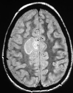
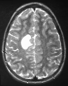
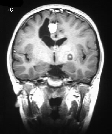
On MR cystic JPA is a well circumscribed mass, hypointense on T1-weighted and hyperintense on T2-weighted images, with a strong enhancing peripheral nodule. Optochiasmatichypothalamic JPA present as multilobulated suprasellar masses, with solid structure. They exhibit low signal on T1-weighted images, high signal on T2-weighted images and variable enhancement. Brain stem lesions cause expansion of the pons or medulla; they are usually solid and appear iso-hypointense on T1-weighted and hyperintense on T2-weighted images. Enhancement is variable and irregular.
Introduction
also called juvenile pilocytic astrocytoma (JPA), a subtype of astrocytoma biologically different from and with a better prognosis than infiltrating astrocytomas. In the WHO classification they are of grade I. The survival rate at 5 years ranges between 80 and 100%.
Age group
JPAs are typically found in children and young adults (35% of all paediatric gliomas).
Location
The most common location is the optic nerves, chiasm and hypothalamus, followed by the cerebellar vermis and hemispheres, brain stem and basal ganglia.
Pathology
Location
The most common location is the optic nerves, chiasm and hypothalamus, followed by the cerebellar vermis and hemispheres, brain stem and basal ganglia.
Pathology
Their gross appearance varies with location: cerebellar astrocytomas are typically cystic with a mural nodule; optic chiasmatic and hypothalamic lesions are solid, multilobulated and well-defined; brain stem astrocytomas appear more diffuse, noninfiltrating lesions.
Clinical features
Clinical features
The clinical presentation varies with location including vomiting, cerebellar ataxia, visual deficits, multiple cranial nerve palsies and sensory-motor disturbances.Obstructive hydrocephalus can be produced by cerebellar astrocytomas


