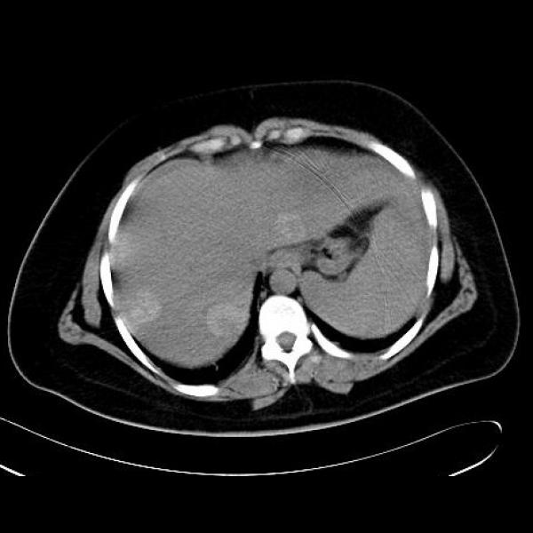
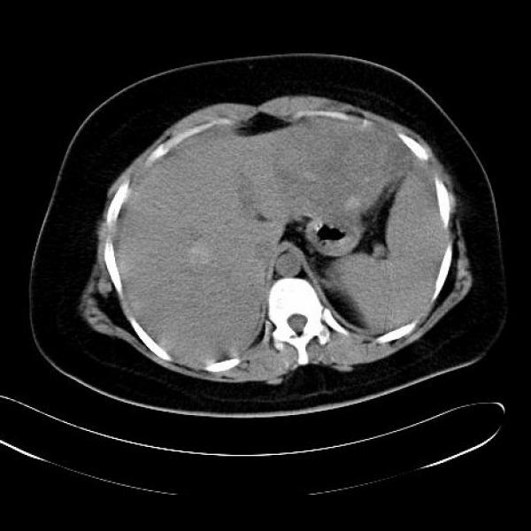
METASTASES FROM MELANOMA
Multiple, well-defined nodular lesions are present in the liver. These are hyperdense on plain CT scan and hyperintense on precontrast T1WI.
Differential diagnosis of T1 hyperintense lesions in the liver:
- Metastases from melanoma
- Metastases fromchoriocarcinoma (with bleed)
- Hepatic adenomas
- Haemorrhagic hepatic cysts
- Dysplastic / regenerative nodules
- Steatosis
- Focal nodular hyperplasia
- Hepatic angiomyolipoma
- Peliosis hepatis
- Lipoma / liposarcoma / pseudolipoma / xanthoma
Substances that cause T1 shortening (hyperintense on T1WI) are:
- Some stages of blood (extracellular methhemoglobin)
- Melanin
- Fat
- Proteinaceous / colloidal material
- Manganese (deposited in the basal ganglia in patients with cirrhosis)
- Gadolinium


