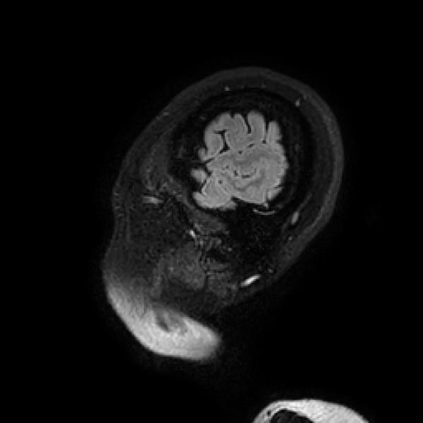
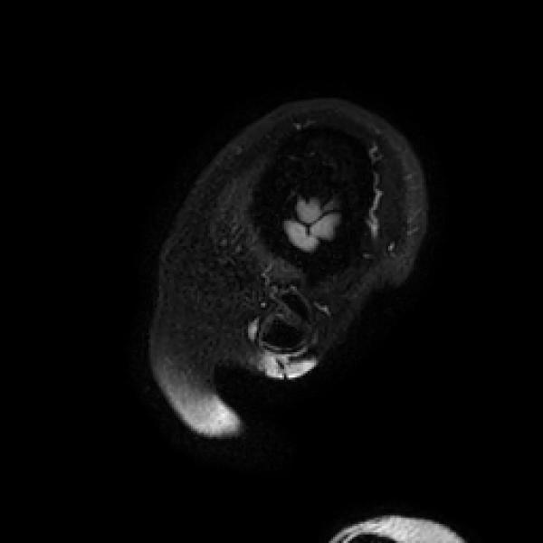
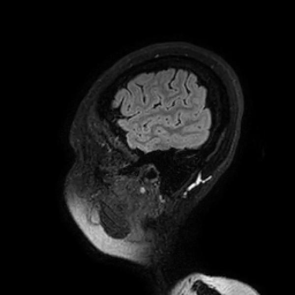
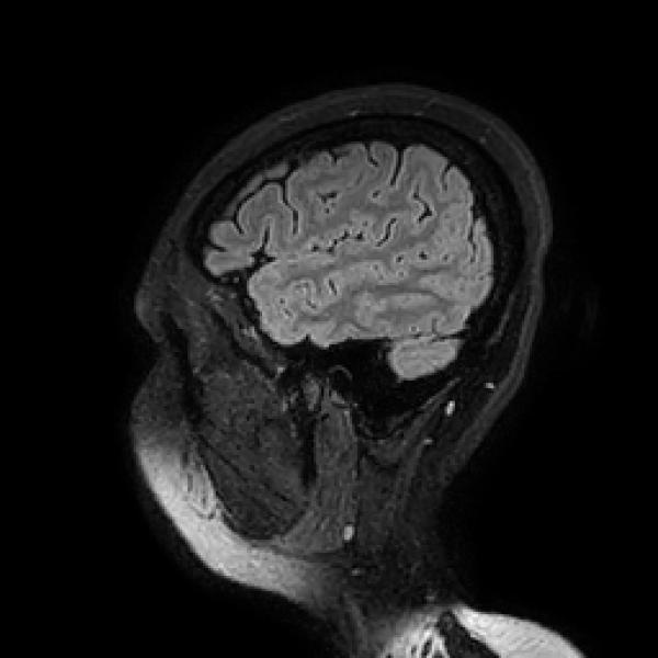
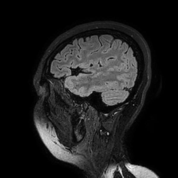
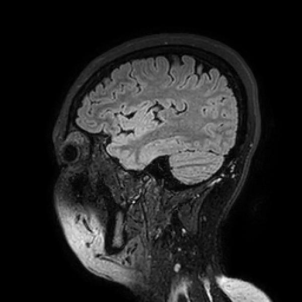
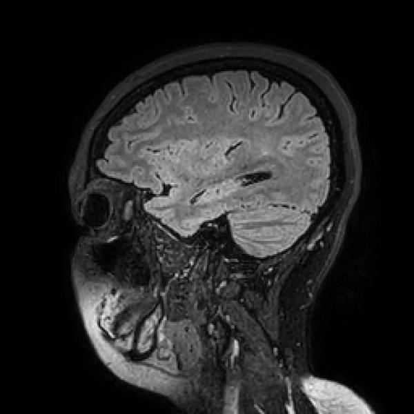
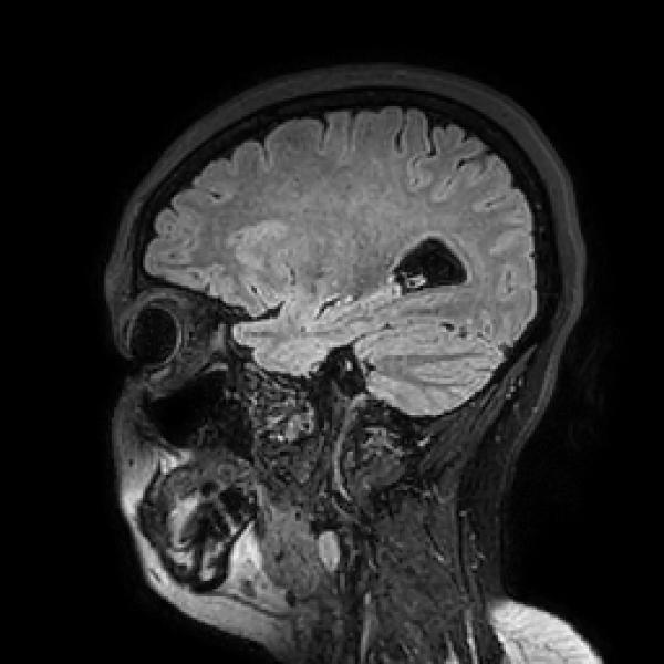
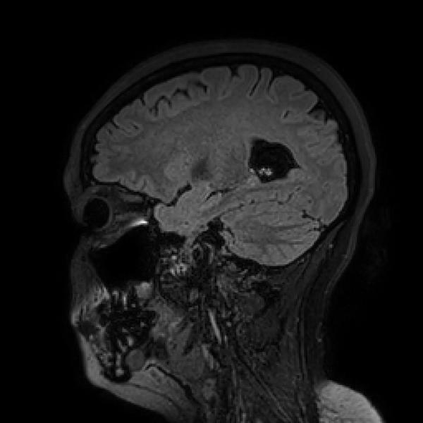
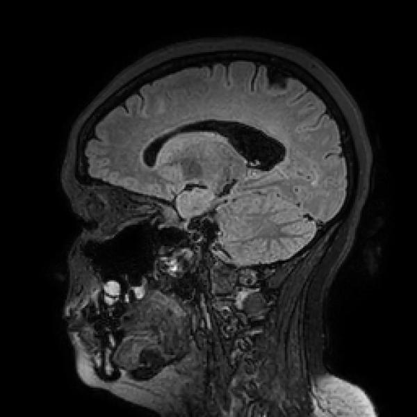
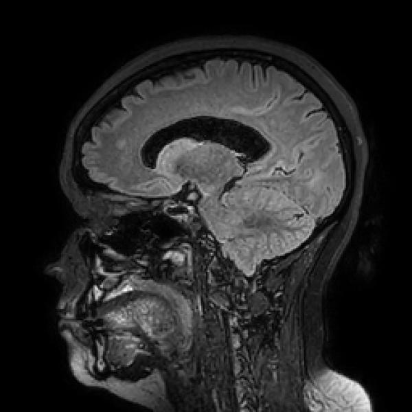
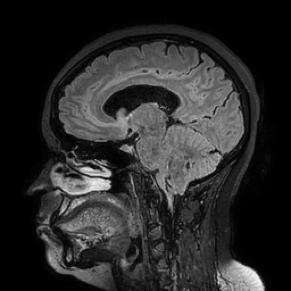
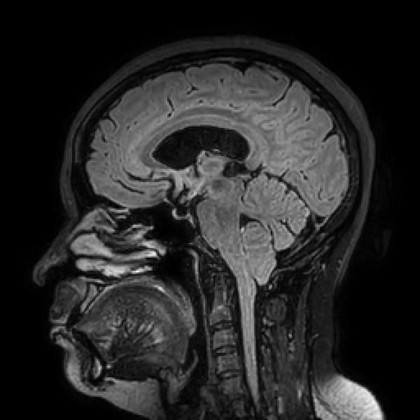
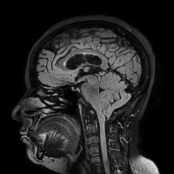
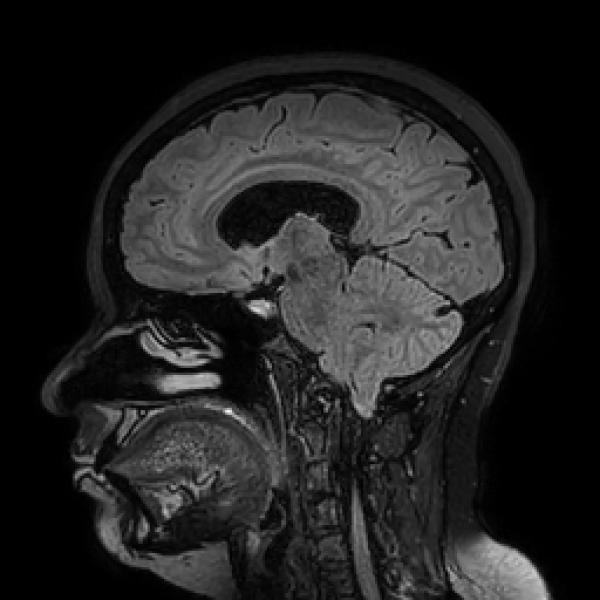
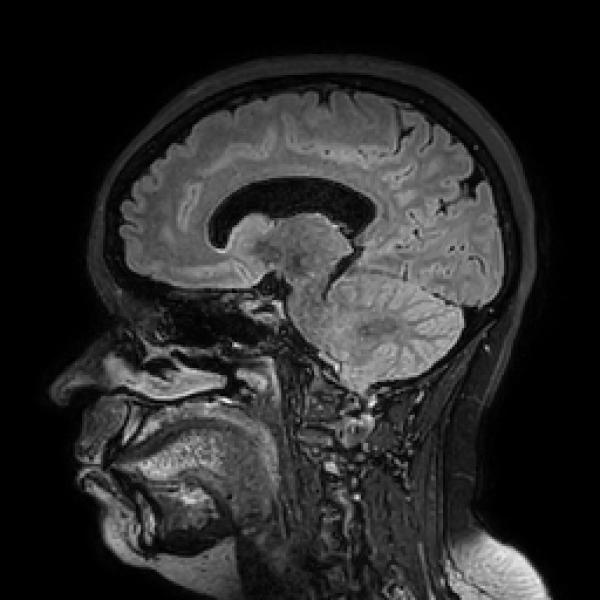
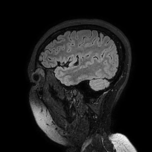
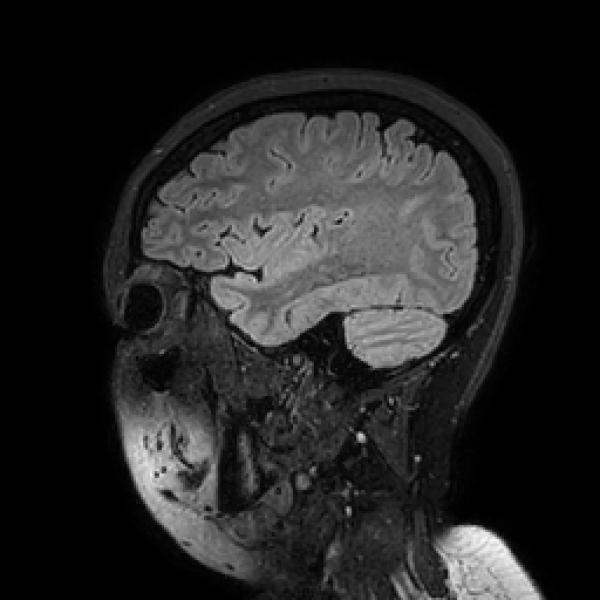
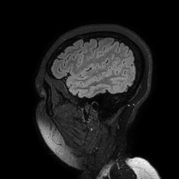
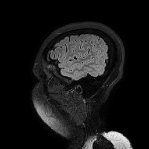
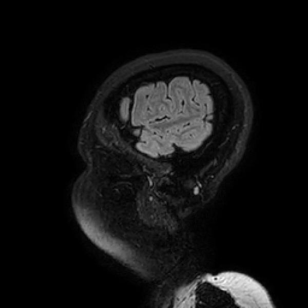
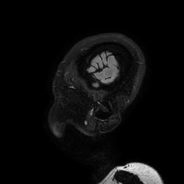
Protocol: Multiplanar multisequence MR imaging.
OBSERVATIONS:
INFRATENTORIAL REGION:
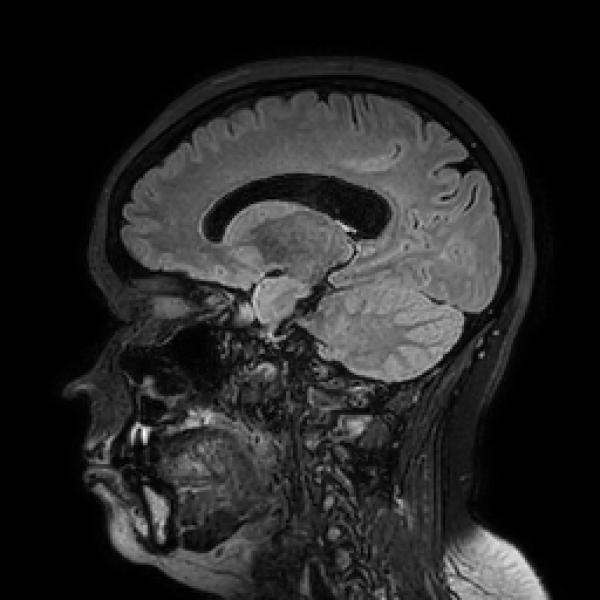
Cerebellar tonsillar herniation is noted ~10mm inferior to the level of foramen magnum.
Cerebellar hemispheres are normal. Fourth ventricle is normal. Brain stem is normal.
Cerebello-pontine angle cisterns are normal.
SUPRATENTORIAL REGION:
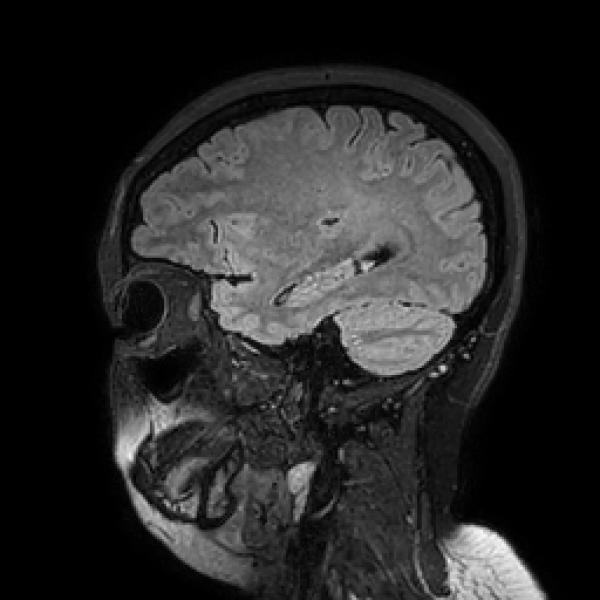
Brain parenchyma shows normal signal intensity. No focal mass lesion.
No evidence of any obvious acute infarct or intracranial haemorrhage.
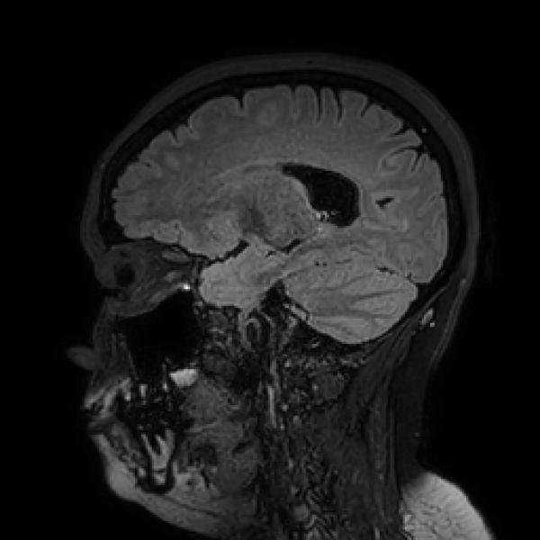
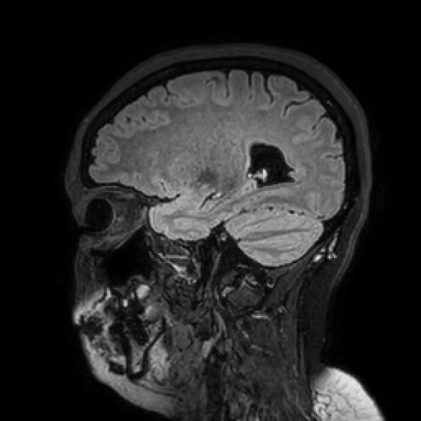
Mild prominence of both lateral ventricles noted. No evidence of periventricular seepage of CSF.
No midline shift.
Cerebral sulci and sylvian fissures are normal. Basal cisterns are normal.
Pituitary gland is flattened along the floor of sella – Consistent with partial empty sella status.
Parasellar regions are normal.
Mild mucosal thickening noted in left maxillary sinus.
Visualized other paranasal sinuses and orbits appear normal.
IMPRESSION:
Ø Mild cerebellar tonsillar herniation – Arnold Chiari Malformation type I.
Ø Mild prominence of both lateral ventricles. No evidence of periventricular seepage of CSF.
No other obvious abnormality detected in the Brain by MR imaging


