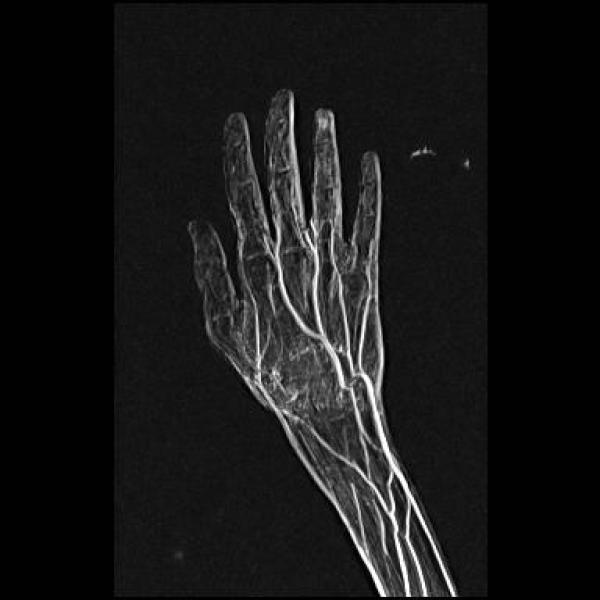
The glomus tumour is one of characteristic fingertip masses that may be diagnosed with MR imaging. The glomus tumour is a hamartoma that arises from the glomus body, an arteriovenous shunt within the dermis that contributes to temperature regulation of the fingers.
The classic clinical presentation of glomus tumour is a triad of fingertip pain, tenderness and sensitivity to pain.
It commonly affects adults and is more common in females than males.
There are two clinical tests which can be used to diagnose glomus tumour.
Hildreth sign, which is disappearance of pain after application of tourniquet proximally on the arm.
Love test, which consist of eliciting pain by applying pressure to precise area with a pencil tip.
MR appearance
With proper MR technique, lesions less than 2mm in size can be detected and localized.
On T1W images, glomus tumour has variable signal characteristics, ranging from low in signal intensity to moderately hyperintense. Hyperintesity is due to hemorrhage within the tumour or due to the large vascular channels within the mass itself. These tumours are highly vascular and shows intense post contrast enhancement.
The high vascularity also accounts for uniform hyperintense signals of these lesions on T2W images.
Contrast enhanced MR angiography can be used in these patients to demonstrate the characteristic vascular nidus found within these lesions


